Sweets, Snacks, and the New Better-for-You Mood
July 3, 2026
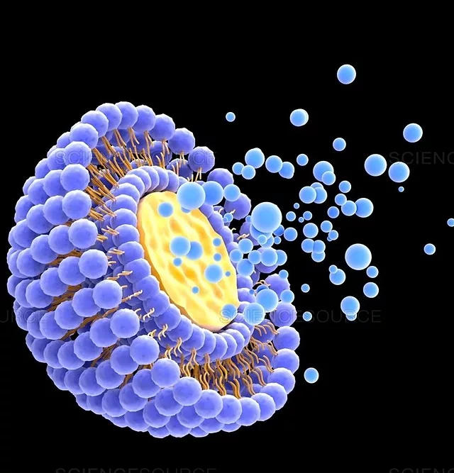
 However, the efficacy of nutraceuticals is a function of absorption and bioavailability after ingestion. Absorption refers to the process of a molecule’s movement from ingestion to its entry into systemic circulation. Bioavailability refers to the amount of the molecule that was absorbed and reached systemic circulation relative to what was ingested.